Category:Chorion
Jump to navigation
Jump to search
protective, noncellular membrane that surrounds the eggs of various animals including insects and fish 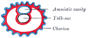 Diagrama mostrant el primer estadi observat d'un embrió humà. | |||||
| Upload media | |||||
| Instance of |
| ||||
|---|---|---|---|---|---|
| Subclass of |
| ||||
| |||||
Media in category "Chorion"
The following 47 files are in this category, out of 47 total.
-
A treatise on the science and practice of midwifery (1880) (14577600379).jpg 780 × 1,460; 239 KB
-
-
Amnion formation in mouse embryos, illustrated by longitudinal sections.jpg 1,200 × 1,394; 1.37 MB
-
Amnion Formation In Mouse Embryos, Illustrated By Transverse Sections.jpg 1,200 × 1,416; 969 KB
-
Amniote embryo ku.jpg 1,162 × 871; 206 KB
-
Amniote embryo.jpg 1,268 × 954; 415 KB
-
Cell populations from chorionic plate and chorionic villi.jpg 955 × 2,083; 274 KB
-
-
Development of the Alimentary Canal, as seen in a Human Embryo about Five Weeks Old.jpg 1,274 × 1,012; 1.5 MB
-
Development of the amnion and allantois.jpg 676 × 902; 1.05 MB
-
EmbryogenesisDynol.svg 512 × 384; 322 KB
-
Embryonic and extraembryonic ectoderm demarcation in the amniochorionic fold.jpg 1,200 × 1,937; 1.62 MB
-
Fetus chorion transverse.jpg 725 × 536; 73 KB
-
Fetus chorion.jpg 453 × 539; 37 KB
-
Foetus cat.jpg 667 × 827; 532 KB
-
Formation of the Umbilicus and Allantois. human embryo, 0.7 mm. long..jpg 1,152 × 803; 970 KB
-
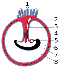
Gray21.png 500 × 427; 24 KB
-
Gray24.png 294 × 127; 5 KB
-
Gray24.svg 252 × 161; 8 KB
-
Gray25.svg 300 × 295; 13 KB
-
Gray26.png 300 × 334; 9 KB
-
Gray26.svg 344 × 345; 21 KB
-
Gray27.png 300 × 298; 10 KB
-
Gray28.svg 396 × 455; 14 KB
-
Gray30.png 500 × 537; 74 KB
-
Gray39.png 500 × 384; 41 KB
-
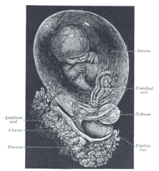
Hand-book of physiology (1892) (14785304043).jpg 1,792 × 888; 162 KB
-
Human embryo Section of embryonic rudiment in Peters' ovum (first week).jpg 1,141 × 857; 540 KB
-
Human- Embryo, about 3.5 mm. long.jpg 803 × 791; 701 KB
-
HumanEmbryogenesis gl.svg 1,560 × 1,220; 686 KB
-
HumanEmbryogenesis ku.svg 1,560 × 1,220; 687 KB
-
HumanEmbryogenesis.svg 1,560 × 1,220; 684 KB
-
-
Morphological differences between human and mouse gastrulation.jpg 2,994 × 3,411; 397 KB
-
Reconstruction of embryos prepared for kaufman's the atlas of mouse development.jpg 1,220 × 2,124; 1.09 MB
-
Sagittal Section of Human Zygote.jpg 817 × 684; 653 KB
-
Schema of a Longitudinal Section of a Human Embryo.jpg 1,125 × 694; 1.02 MB
-
-
-
-
Series of longitudinal sections of an embryo with large exocoelomic cavity (ec).jpg 1,220 × 1,217; 1.12 MB
-
-
Structure of the human amniotic membrane.jpg 3,313 × 3,966; 832 KB
-
-
Ultrasound Scan ND 034.jpg 491 × 396; 49 KB
-
Umbilical Region of a Human Embryo 10 mm. in length.jpg 1,167 × 957; 1.09 MB
-
Yolk sacs.png 1,346 × 511; 294 KB