Category:Breast cancer
Zur Navigation springen
Zur Suche springen
Radiologie: Ultraschall · Röntgenstrahlung · Computertomographie · Magnetic resonance · Positronen-Emissions-Tomographie · Skelettszintigrafie | Anatomische Pathologie: Cytopathologie · Gross pathology · Histopathologie | Othering: Epidemiologie (Karte → Welt) · Stadienbestimmung | Dateiformat: Scalable Vector Graphics · Videotechnik | 
bösartiger Tumor der Brustdrüse des Menschen  Imatge histopatològica del carcinoma de cèl·lules ductals in situ (DCIS) de mama. Taca d'hematoxilina-eosina. | |||||
| Medium hochladen | |||||
| Audiodatei eines gesprochenen Texts | |||||
|---|---|---|---|---|---|
| Ist ein(e) |
| ||||
| Unterklasse von |
| ||||
| Aspekt von | |||||
| |||||
Unterkategorien
Es werden 39 von insgesamt 39 Unterkategorien in dieser Kategorie angezeigt:
In Klammern die Anzahl der enthaltenen Kategorien (K), Seiten (S), Dateien (D)
- PET images of breast cancer (4 D)
- Videos of breast cancer (145 D)
A
B
- Breast biopsy (4 D)
- Breast self-examinations (34 D)
- BrM2 cells (3 D)
C
D
E
- Experimental mammary neoplasms (18 D)
F
- Betty Ford and breast cancer (25 D)
I
M
- MCF-7 cells (93 D)
- Media from NPJ breast cancer (5 D)
O
R
S
- Sacituzumab govitecan (2 D)
- Sentinel lymph node biopsy (5 D)
Medien in der Kategorie „Breast cancer“
Folgende 200 Dateien sind in dieser Kategorie, von 205 insgesamt.
(vorherige Seite) (nächste Seite)-
1 applicator.jpg 2.592 × 1.944; 1,88 MB
-
Tumors of the female breast - a clinical lecture (IA 101726975.nlm.nih.gov).pdf 595 × 947, 20 Seiten; 517 KB
-
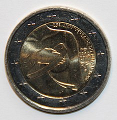
2 Euro - 25 Jahre Rosa Schleife – Kampf gegen den Brustkrebs.jpg 1.848 × 1.892; 1,3 MB
-
3D Dual Color Super Resolution Microscopy Cremer 2010.png 3.486 × 1.280; 3 MB
-
3D medical animation TNM Staging System.jpg 1.920 × 1.080; 1.002 KB
-
-
-
-
All women need to know about breat cancer and early detection exhibit.jpg 2.704 × 2.193; 3,96 MB
-
Amino acid metabolism in triple-negative breast cancer cells.svg 1.424 × 982; 101 KB
-
-
Antiestrogen.png 1.974 × 1.092; 782 KB
-
Areola Micropigmentation.jpg 3.088 × 2.320; 262 KB
-
Arimidex.jpg 2.115 × 2.182; 3,23 MB
-
Aromatase Inhibitor.svg 512 × 384; 604 KB
-
Autobus rabat maroc cancer sein prévention.jpg 4.288 × 2.848; 6,28 MB
-
Bc-classification.png 1.000 × 1.500; 1,29 MB
-
Bc-ica-colour.png 550 × 550; 62 KB
-
Bc-isomap-colour.png 550 × 550; 102 KB
-
Bc-mfa-allgene-colour.png 550 × 550; 63 KB
-
Bc-mfa-nfkbpathway-colour.png 550 × 550; 63 KB
-
Bc-mfa-rbpathway-colour.png 550 × 550; 53 KB
-
Bc-mfa-tgfbetapathway-colour.png 550 × 550; 66 KB
-
Bc-nmf-colour.png 550 × 550; 66 KB
-
Bc-pca-colour.png 550 × 550; 72 KB
-
Bc-subtypes.png 550 × 550; 60 KB
-
FEDLINK - United States Federal Collection (IA billtoamendtitle00uni z8b).pdf 552 × 829, 8 Seiten; 303 KB
-
Bio-Assembler vs. Matrigel with Human Mammary Epithelial Cells.tiff 690 × 536; 109 KB
-
BIRADS Category III Lesion.svg 1.567 × 411; 95 KB
-
Blood test kit (4141300940).jpg 2.048 × 1.536; 1,45 MB
-
BRCA .tif 970 × 658; 274 KB
-
BRCA1 and BRCA2 mutations and absolute cancer risk (Updated version- 2023).jpg 1.920 × 1.197; 199 KB
-
BRCA1 and BRCA2 mutations and absolute cancer risk (Updated-2023).jpg 1.949 × 623; 244 KB
-
BRCA1MutIdeogram.png 630 × 560; 80 KB
-
Breast cancer acinar structures cultured by MLM.tif 717 × 496; 2,57 MB
-
Breast cancer cell (2).jpg 1.800 × 1.844; 642 KB
-
Breast cancer cells.tif 512 × 512; 770 KB
-
Breast cancer grafts updated.jpg 595 × 348; 27 KB
-
Breast cancer grafts-4.jpg 595 × 348; 28 KB
-
Breast cancer grafts-6.jpg 615 × 348; 28 KB
-
Breast cancer grafts.jpg 576 × 348; 25 KB
-
Breast cancer gross appearance.jpg 400 × 300; 135 KB
-
Breast cancer illustrations-ar.jpg 317 × 464; 29 KB
-
Breast cancer image.webp 1.100 × 560; 247 KB
-
Breast cancer incidence by age.tif 1.314 × 994; 181 KB
-
Breast cancer incidence by anatomical site (females)-ar.png 650 × 587; 72 KB
-
Breast Cancer mice.jpg 833 × 657; 88 KB
-
Breast cancer mice.jpg 4.224 × 2.368; 2,78 MB
-
Breast cancer probability according to mammography.svg 989 × 609; 17 KB
-
Breast cancer progression.svg 488 × 135; 233 KB
-
Breast cancer sample, being subjected to FISH analysis.jpg 184 × 164; 3 KB
-
Breast cancer sensitization program.jpg 1.032 × 726; 293 KB
-
Breast cancer specimen.jpg 576 × 1.024; 96 KB
-
Breast cancer spheroids with aptamers.png 728 × 726; 613 KB
-
-
Breast Cancer-ar.png 480 × 600; 271 KB
-
Breast Cancer.png 959 × 1.086; 712 KB
-
Breast dce-mri.jpg 378 × 297; 50 KB
-
Breast Implants & Cancer (5387274671)-ar.jpg 800 × 600; 615 KB
-
Breast Implants & Cancer (5387274671).jpg 3.000 × 2.250; 1,86 MB
-
Breast Cancer in North Carolina- A Handbook for Health Care Providers (IA breastcancerinno00nort).pdf 1.468 × 2.183, 68 Seiten; 3,4 MB
-
Breast Cancer- A Resource Guide for Minority Women (IA breastcancerreso00offi).pdf 843 × 1.145, 32 Seiten; 2,54 MB
-
Breast Cancer Resource Guide for Minority Women (IA breastcancerreso00unse).pdf 1.241 × 1.597, 36 Seiten; 2,17 MB
-
Breast Cancer- A Resource for Minority Women (IA breastcancerreso00unse 0).pdf 1.504 × 1.931, 64 Seiten; 2,3 MB
-
BreastCancerRightSamplel.jpg 512 × 512; 113 KB
-
Breast and Cervical Cancer Screening- Barriers and Use Among Specific Populations, Supplement 3 (IA breastcervicalca00case).pdf 1.406 × 1.943, 58 Seiten; 2,61 MB
-
-
-
Brust-epithese-rueckseite.jpg 3.024 × 3.024; 1,4 MB
-
Brust-epithese-vorne-glatt.jpg 3.024 × 3.024; 1,67 MB
-
Brust-epithese-vorne.jpg 3.024 × 3.024; 1,03 MB
-
Brust.png 309 × 285; 15 KB
-
Cancerdusein2 Wiki.jpg 500 × 480; 89 KB
-
Cancro da Mama.jpg 990 × 556; 82 KB
-
Cannon ball mets in ca breast.jpg 1.277 × 633; 96 KB
-
Carcinomatous growth on the breast Wellcome L0062165.jpg 6.048 × 4.528; 5,73 MB
-
Case TCGA-AN-A046 slide 01Z-00-DX1 from the TCGA-BRCA project.png 3.072 × 1.712; 2,86 MB
-
Cathrin Brisken Portrait 2006.jpg 4.368 × 2.912; 1,96 MB
-
Ccis rate and bc.webp 410 × 244; 7 KB
-
Cecilie - Breast Cancer Statue.jpg 2.268 × 3.792; 7,12 MB
-
Cell morphology and mechanics are vimentin dependent.jpg 1.612 × 1.150; 748 KB
-
Cellular adhesion on submicron topographies controls EMTMET.jpg 916 × 1.840; 891 KB
-
CeRNA counteracting miRNA negative regulation of PTEN.jpg 1.404 × 1.524; 175 KB
-
Chemotherapy with acral cooling.jpg 1.200 × 1.600; 336 KB
-
-
-
-
Connie Johnson (cropped).jpg 260 × 320; 22 KB
-
Current multidisciplinary treatment of TNBC.webp 685 × 318; 28 KB
-
Developing evidence-based measures of care for breast cancer (IA developingeviden00pear).pdf 629 × 827, 210 Seiten; 6,66 MB
-
Diagnostics-09-00012-g011.jpg 737 × 442; 91 KB
-
Diagnostika-raka 4.jpg 638 × 443; 34 KB
-
-
Diffuse Optical Tomography workflow - journal.pone.0045714.g001.png 2.704 × 1.887; 773 KB
-
Distribución cancer de mama.svg 495 × 350; 14 KB
-
DSLRF image.jpg 300 × 119; 50 KB
-
-
-
Epitope-mapping illustration-6-copy.png 1.309 × 855; 456 KB
-
-
-
-
-
-
Estrogenic endocrine disruptors by Marta.jpg 628 × 413; 104 KB
-
Events hall.jpg 640 × 480; 158 KB
-
Expression 2.png 788 × 592; 67 KB
-
-
Fashion Parade Breast Cancer Survivors.jpg 3.014 × 2.009; 4,88 MB
-
Figure 1 (11108271853).png 444 × 349; 327 KB
-
Figure 1 (7769085132).png 636 × 909; 938 KB
-
Figure 4B (6983906744).png 887 × 695; 956 KB
-
Figure S2 (colour inverted) (8007511032).png 801 × 853; 1,81 MB
-
Figure S3.6 (8165172983).png 724 × 698; 1,2 MB
-
Figure S3.7 (7876918270).png 661 × 912; 1,16 MB
-
Firma de reordenamiento.png 878 × 748; 211 KB
-
Firma de sustitución de bases.jpg 596 × 503; 30 KB
-
FISH del gen Her2, detalle.jpg 1.920 × 1.036; 79 KB
-
FISH del gen Her2.jpg 1.920 × 1.036; 130 KB
-
Five year survival rates from breast cancer, OWID.svg 850 × 600; 141 KB
-
From Dallas Terre Quinn operates and uplifts..jpg 1.080 × 2.340; 520 KB
-
Gail Lebovic at School of Oncoplastic Surgery.jpg 2.800 × 1.867; 3,61 MB
-
-
Handwritten subject card..JPG 768 × 526; 200 KB
-
HER2 SISH demonstrating HER2 amplification in male breast cancer.jpg 673 × 536; 262 KB
-
Her2neu 3+staining.jpg 4.912 × 3.684; 3,68 MB
-
Heterogeneous breast tumor in 3D.jpg 2.048 × 2.048; 1,78 MB
-
Hibridación in situ del gen Her2 (no amplificado).jpg 1.920 × 1.036; 165 KB
-
Hibridación in situ fluorescente (FISH) del Gen Her2 (no amplificado) (mama) (05).jpg 1.920 × 1.036; 239 KB
-
-
-
-
-
-
Hibridación in situ fluorescente del gen Her2 (FISH del gen Her2; 5 señales).jpg 1.920 × 1.036; 227 KB
-
Hy-Կրծքագեղձի քաղցկեղ (Breast cancer).ogg 4 min 18 s; 9,92 MB
-
ICI7320 Breast exam.jpg 255 × 223; 12 KB
-
Immunohistochemical staining of male breast cancer for ER and PgR.jpg 758 × 546; 298 KB
-
Inherited breast cancer es.svg 300 × 270; 262 KB
-
International day against breast cancer in Didouche mourad, Algiers.jpg 1.706 × 1.280; 538 KB
-
Invasive Breast Cancer in 3D.jpg 1.557 × 1.296; 597 KB
-
Invasive ductal carcinoma.jpg 4.096 × 3.008; 2,38 MB
-
Invasive lobular carcinoma.jpg 4.096 × 3.008; 2,58 MB
-
Jean Doerge Reunio281.jpg 720 × 540; 53 KB
-
Kataegi.png 340 × 278; 23 KB
-
Key signaling pathways in breast cancer.webp 2.000 × 1.027; 203 KB
-
Laura Bush meets with women in UAE.jpg 514 × 343; 66 KB
-
Main features of breast cancer molecular subtypes.webp 410 × 223; 10 KB
-
-
Mammella cancro2.jpg 640 × 480; 109 KB
-
MBq-Sono-Brustkrebs.jpg 569 × 379; 22 KB
-
MCF-7 Cells.jpg 1.600 × 1.200; 723 KB
-
Measurement of tumor size on two microscopy slides.jpg 1.601 × 1.401; 544 KB
-
Breast Cancer- Know the Facts - A Situation No Woman Wants To Face! (IA MH95D2488).pdf 883 × 2.127, 8 Seiten; 243 KB
-
Miss Mosley, afflicted with breast cancer Wellcome L0049815.jpg 5.955 × 4.367; 3,58 MB
-
Modern surgery, general and operative (1914) (14596177937).jpg 1.202 × 1.066; 184 KB
-
Mrs Broadbent, afflicted with breast cancer Wellcome L0049816.jpg 4.687 × 5.618; 3,76 MB
-
Multicultural Aspects of Breast Cancer Etiology (IA multiculturalasp00nati).pdf 1.200 × 1.606, 88 Seiten; 4,72 MB
-
Mutação dos genes BRCA1 e BRCA2 de portadoras de câncer de mama.png 628 × 439; 86 KB
-
Nachsorgepass Brustkrebs.jpg 1.135 × 940; 133 KB
-
Nachsorgepass Brustkrebs1.jpg 1.148 × 921; 155 KB
-
National Plan of Action on Asian American Women and Breast Cancer (IA nationalplanofac00nati).pdf 1.200 × 1.825, 36 Seiten; 1,78 MB
-
Native Women's Breast and Cervical Health Magazine (IA nativewomensbrea00prov).pdf 1.489 × 1.908, 36 Seiten; 4,32 MB
-
NCBI Geo Profile for Triple Negative Breast Cancer and YPLR6490.png 594 × 358; 86 KB
-
Neglected breast cancer (Cancer du sein négligé).jpg 960 × 720; 97 KB
-
Neglected breast cancer.jpg 960 × 720; 102 KB
-
Neuropilin-2 (Nrp2) expression in normal breast and breast carcinoma tissue.jpg 1.200 × 900; 498 KB
-
Nipple Aspirate Test No Substitute for Mammogram (11342754456).jpg 2.655 × 5.041; 2,16 MB
-
Nolvadex.jpg 2.119 × 1.423; 574 KB
-
Octobre-rose.jpg 1.010 × 1.769; 641 KB
-
-
Patricia JANEČKOVÁ - For all my fans.webm 1 min 3 s, 1.920 × 1.080; 12,61 MB
-
PBB Protein BRCA2 image.jpg 500 × 500; 30 KB
-
Prior mammography utilization - does it explain black-white differences in breast cancer outcomes? (IA priormammography00mcca).pdf 1.204 × 1.620, 218 Seiten; 5,08 MB
-
Rainplot for Kataegis in Breast Cancer Genome.jpg 2.482 × 1.878; 1,06 MB
-
Rakukultuuri kasvatamine.jpg 2.848 × 2.136; 471 KB
-
-
Relevant molecular pathways and targeted agents in TNBC.jpg 728 × 850; 138 KB
-
Samuel and Connie Johnson (cropped).jpg 216 × 314; 21 KB
-
Samuel and Connie Johnson.jpg 1.024 × 683; 73 KB
-
Savethenipple post ENG1.jpg 1.400 × 811; 517 KB
-
Schematic representation of breast cancer metastatic study models.pdf 1.650 × 1.275; 704 KB
-
Scheme BRCA.png 1.154 × 644; 69 KB
-
Schéma-du-sein.jpg 497 × 350; 96 KB
-
Sister two.jpg 200 × 223; 22 KB
-
Slow motion arrows.gif 300 × 247; 12,81 MB
-
Stage 4 of Breast Cancer.jpg 1.920 × 1.080; 917 KB
-
-
Tamoxifen and breast cancer.jpg 1.449 × 1.147; 1,05 MB
-
Technician performing laboratory test.jpg 2.700 × 1.800; 2,01 MB
-
The Anastacia Fund logo.jpg 250 × 164; 16 KB
-
The bioabsorbable 3D marker with clips embedded.jpg 1.224 × 1.632; 278 KB
-
-
The structure of the mammary gland and the origin of BC cells.webp 1.944 × 885; 203 KB
-
Tissue culture hood.png 428 × 310; 245 KB
-
Top Four Mammogram Myths (8726196704).jpg 2.658 × 7.753; 3,96 MB
-
-
Unidad móvil de detección precoz cáncer de mama (37090066122).jpg 4.608 × 3.072; 7,28 MB
-
Version2 savethenipple post ENG3.jpg 1.400 × 811; 489 KB
-
Vestito di cellule.jpg 768 × 768; 31 KB