Corticopontine fibers: Difference between revisions
Content deleted Content added
removed Category:Neuroanatomy; added Category:Cerebral white matter using HotCat |
m Replace magic links with templates per local RfC and MediaWiki RfC |
||
| Line 17: | Line 17: | ||
| BrainInfoNumber = 375 |
| BrainInfoNumber = 375 |
||
}} |
}} |
||
'''Corticopontine fibers''' <ref>Kamali A, Kramer LA, Frye RE, Butler IJ, Hasan KM. Diffusion tensor tractography of the human brain cortico-ponto-cerebellar pathways: a quantitative preliminary study. J Magn Reson Imaging. 2010 Oct;32(4):809-17. doi: 10.1002/jmri.22330. PMID |
'''Corticopontine fibers''' <ref>Kamali A, Kramer LA, Frye RE, Butler IJ, Hasan KM. Diffusion tensor tractography of the human brain cortico-ponto-cerebellar pathways: a quantitative preliminary study. J Magn Reson Imaging. 2010 Oct;32(4):809-17. doi: 10.1002/jmri.22330. {{PMID|20882611}} |
||
</ref> are projections from the [[cerebral cortex]] to the [[pontine nuclei]].<ref name="pmid18982130">{{cite journal |author=Leergaard TB, Bjaalie JG |title=Topography of the complete corticopontine projection: From experiments to principal Maps |journal=Front Neurosci |volume=1 |issue=1 |pages=211–23 |date=November 2007 |pmid=18982130 |pmc=2518056 |doi=10.3389/neuro.01.1.1.016.2007 }}</ref> |
</ref> are projections from the [[cerebral cortex]] to the [[pontine nuclei]].<ref name="pmid18982130">{{cite journal |author=Leergaard TB, Bjaalie JG |title=Topography of the complete corticopontine projection: From experiments to principal Maps |journal=Front Neurosci |volume=1 |issue=1 |pages=211–23 |date=November 2007 |pmid=18982130 |pmc=2518056 |doi=10.3389/neuro.01.1.1.016.2007 }}</ref> |
||
Revision as of 15:15, 22 May 2017
This article needs additional citations for verification. (November 2008) |
| Corticopontine fibers | |
|---|---|
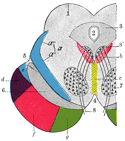 Coronal section through mid-brain. 1. Corpora quadrigemina. 2. Cerebral aqueduct. 3. Central gray stratum. 4. Interpeduncular space. 5. Sulcus lateralis. 6. Substantia nigra. 7. Red nucleus of tegmentum. 8. Oculomotor nerve, with 8’, its nucleus of origin. a. Lemniscus (in blue) with a’ the medial lemniscus and a" the lateral lemniscus. b. Medial longitudinal fasciculus. c. Raphé. d. Temporopontine fibers. e. Portion of medial lemniscus, which runs to the lentiform nucleus and insula. f. Cerebrospinal fibers. g. Frontopontine fibers. | |
| Details | |
| Identifiers | |
| Latin | fibrae corticopontinae, tractus corticopontinus |
| NeuroNames | 1322 |
| TA98 | A14.1.05.107 |
| TA2 | 5619 |
| FMA | 75190 |
| Anatomical terms of neuroanatomy | |
Corticopontine fibers [1] are projections from the cerebral cortex to the pontine nuclei.[2]
Depending upon the lobe of origin, they can be classified as frontopontine fibers, parietopontine fibers, temporopontine fibers or occipitopontine fibers.[3]
References
- ^ Kamali A, Kramer LA, Frye RE, Butler IJ, Hasan KM. Diffusion tensor tractography of the human brain cortico-ponto-cerebellar pathways: a quantitative preliminary study. J Magn Reson Imaging. 2010 Oct;32(4):809-17. doi: 10.1002/jmri.22330. PMID 20882611
- ^ Leergaard TB, Bjaalie JG (November 2007). "Topography of the complete corticopontine projection: From experiments to principal Maps". Front Neurosci. 1 (1): 211–23. doi:10.3389/neuro.01.1.1.016.2007. PMC 2518056. PMID 18982130.
{{cite journal}}: CS1 maint: unflagged free DOI (link) - ^ http://braininfo.rprc.washington.edu/AncilDefinition.aspx?ID=1322&questID=1322
External links
This neuroanatomy article is a stub. You can help Wikipedia by expanding it. |