Corticopontine fibers: Difference between revisions
Content deleted Content added
Tom.Reding (talk | contribs) m +{{Authority control}} (1 source from Wikidata), WP:GenFixes on, |
Fixed link #article-section-source-editor Tags: Mobile edit Mobile app edit iOS app edit |
||
| (4 intermediate revisions by 4 users not shown) | |||
| Line 1: | Line 1: | ||
{{Short description|Projections from the cerebral cortex to the pontine nuclei}} |
|||
{{Use American English|date=March 2021}} |
|||
{{Use mdy dates|date=March 2021}} |
|||
{{More citations needed|date=November 2008}} |
{{More citations needed|date=November 2008}} |
||
{{Infobox brain |
{{Infobox brain |
||
| Line 4: | Line 7: | ||
| Latin = fibrae corticopontinae, tractus corticopontinus |
| Latin = fibrae corticopontinae, tractus corticopontinus |
||
| Image = Gray710.png |
| Image = Gray710.png |
||
| Caption = Coronal section through [[mid-brain]]. <br>1. [[Corpora quadrigemina]]. <br>2. [[Cerebral aqueduct]]. <br>3. [[Central gray stratum]]. <br>4. [[Interpeduncular space]]. <br>5. [[Sulcus lateralis]]. <br>6. [[Substantia nigra]]. <br>7. [[Red nucleus]] of [[tegmentum]]. <br>8. [[Oculomotor nerve]], with 8’, its nucleus of origin. a. [[Lemniscus (anatomy)|Lemniscus]] (in blue) with a’ the [[medial lemniscus]] and a" the [[lateral lemniscus]]. b. [[Medial longitudinal fasciculus]]. c. [[Raphe|Raphé]]. d. Temporopontine fibers. e. Portion of [[medial lemniscus]], which runs to the [[lentiform nucleus]] and [[Insular cortex|insula]]. f. [[Cerebrospinal fibers]]. g. [[Frontopontine fibers]]. |
| Caption = Coronal section through [[mid-brain]]. <br>1. [[Corpora quadrigemina]]. <br>2. [[Cerebral aqueduct]]. <br>3. [[Central gray stratum]]. <br>4. [[Interpeduncular space]]. <br>5. [[Sulcus lateralis]]. <br>6. [[Substantia nigra]]. <br>7. [[Red nucleus]] of [[tegmentum]]. <br>8. [[Oculomotor nerve]], with 8’, its nucleus of origin. a. [[Lemniscus (anatomy)|Lemniscus]] (in blue) with a’ the [[medial lemniscus]] and a" the [[lateral lemniscus]]. b. [[Medial longitudinal fasciculus]]. c. [[Raphe nuclei|Raphé]]. d. Temporopontine fibers. e. Portion of [[medial lemniscus]], which runs to the [[lentiform nucleus]] and [[Insular cortex|insula]]. f. [[Cerebrospinal fibers]]. g. [[Frontopontine fibers]]. |
||
| Image2 = |
| Image2 = |
||
| Caption2 = |
| Caption2 = |
||
| Line 11: | Line 14: | ||
| Vein = |
| Vein = |
||
}} |
}} |
||
'''Corticopontine fibers'''<ref>Kamali A, Kramer LA, Frye RE, Butler IJ, Hasan KM. Diffusion tensor tractography of the human brain cortico-ponto-cerebellar pathways: a quantitative preliminary study. J Magn Reson Imaging. 2010 Oct;32(4):809-17. doi: 10.1002/jmri.22330. {{PMID|20882611}} |
|||
</ref> are projections from the [[cerebral cortex]] to the [[pontine nuclei]].<ref name="pmid18982130">{{cite journal |author=Leergaard TB, Bjaalie JG |title=Topography of the complete corticopontine projection: From experiments to principal Maps |journal=Front Neurosci |volume=1 |issue=1 |pages=211–23 |date=November 2007 |pmid=18982130 |pmc=2518056 |doi=10.3389/neuro.01.1.1.016.2007 }}</ref> |
'''Corticopontine fibers'''<ref>{{cite journal | author = Kamali A, Kramer LA, Frye RE, Butler IJ, Hasan KM | date = Oct 2010 | title = Diffusion tensor tractography of the human brain cortico-ponto-cerebellar pathways: a quantitative preliminary study | journal = J Magn Reson Imaging | volume = 32 | issue = 4| pages = 809–17 | doi = 10.1002/jmri.22330 | pmid = 20882611 | pmc = 4492525 }}</ref> are projections from the [[cerebral cortex]] to the [[pontine nuclei]].<ref name="pmid18982130">{{cite journal |author=Leergaard TB, Bjaalie JG |title=Topography of the complete corticopontine projection: From experiments to principal Maps |journal=Front Neurosci |volume=1 |issue=1 |pages=211–23 |date=November 2007 |pmid=18982130 |pmc=2518056 |doi=10.3389/neuro.01.1.1.016.2007 |doi-access=free }}</ref> |
||
Depending upon the lobe of origin, they can be classified as [[frontopontine fibers]], [[parietopontine fibers]], [[temporopontine fibers]] or [[occipitopontine fibers]].<ref>http://braininfo.rprc.washington.edu/AncilDefinition.aspx?ID=1322&questID=1322{{dead link|date=August 2017 |bot=InternetArchiveBot |fix-attempted=yes }}</ref> |
Depending upon the lobe of origin, they can be classified as [[frontopontine fibers]], [[parietopontine fibers]], [[temporopontine fibers]] or [[occipitopontine fibers]].<ref>http://braininfo.rprc.washington.edu/AncilDefinition.aspx?ID=1322&questID=1322{{dead link|date=August 2017 |bot=InternetArchiveBot |fix-attempted=yes }}</ref> |
||
Latest revision as of 19:10, 23 February 2024
This article needs additional citations for verification. (November 2008) |
| Corticopontine fibers | |
|---|---|
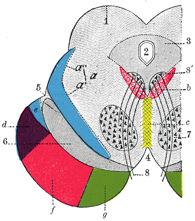 Coronal section through mid-brain. 1. Corpora quadrigemina. 2. Cerebral aqueduct. 3. Central gray stratum. 4. Interpeduncular space. 5. Sulcus lateralis. 6. Substantia nigra. 7. Red nucleus of tegmentum. 8. Oculomotor nerve, with 8’, its nucleus of origin. a. Lemniscus (in blue) with a’ the medial lemniscus and a" the lateral lemniscus. b. Medial longitudinal fasciculus. c. Raphé. d. Temporopontine fibers. e. Portion of medial lemniscus, which runs to the lentiform nucleus and insula. f. Cerebrospinal fibers. g. Frontopontine fibers. | |
| Details | |
| Identifiers | |
| Latin | fibrae corticopontinae, tractus corticopontinus |
| NeuroNames | 1322 |
| TA98 | A14.1.05.107 |
| TA2 | 5619 |
| FMA | 75190 |
| Anatomical terms of neuroanatomy | |
Corticopontine fibers[1] are projections from the cerebral cortex to the pontine nuclei.[2]
Depending upon the lobe of origin, they can be classified as frontopontine fibers, parietopontine fibers, temporopontine fibers or occipitopontine fibers.[3]
References
[edit]- ^ Kamali A, Kramer LA, Frye RE, Butler IJ, Hasan KM (October 2010). "Diffusion tensor tractography of the human brain cortico-ponto-cerebellar pathways: a quantitative preliminary study". J Magn Reson Imaging. 32 (4): 809–17. doi:10.1002/jmri.22330. PMC 4492525. PMID 20882611.
{{cite journal}}: CS1 maint: multiple names: authors list (link) - ^ Leergaard TB, Bjaalie JG (November 2007). "Topography of the complete corticopontine projection: From experiments to principal Maps". Front Neurosci. 1 (1): 211–23. doi:10.3389/neuro.01.1.1.016.2007. PMC 2518056. PMID 18982130.
- ^ http://braininfo.rprc.washington.edu/AncilDefinition.aspx?ID=1322&questID=1322[permanent dead link]
External links
[edit]Cortex->Pons->Cerebellum:
This neuroanatomy article is a stub. You can help Wikipedia by expanding it. |