Nucleoprotein: Difference between revisions
→List of RNPs: examples already present in prose |
→Ribonucleoproteins: updating over-complex caption. Still complex, but reordered to be easier to read. |
||
| Line 14: | Line 14: | ||
== Ribonucleoproteins == |
== Ribonucleoproteins == |
||
[[File:A-Ribonucleoprotein-Complex-Protects-the-Interleukin-6-mRNA-from-Degradation-by-Distinct-ppat.1004899.s011.ogv|thumb| |
[[File:A-Ribonucleoprotein-Complex-Protects-the-Interleukin-6-mRNA-from-Degradation-by-Distinct-ppat.1004899.s011.ogv|thumb|[[Cell nucleus]] with DNA stained blue, and [[Nucleolin|nucleolin protein]] in red. The nucleolin protein binds some [[MRNA|mRNAs]] (e.g. mRNA for [[Interleukin-6]]). This protects those mRNAs from degradation by [[Kaposi's sarcoma-associated herpesvirus]] when infected. This RNA-nucleolin complex is then safely transported to the cytosol for translation by ribosomes to produce the Interleukin-6 protein, which is involved in [[Innate immune system|antiviral immune response]].<ref>{{Cite journal|last=Muller|first=Mandy|last2=Hutin|first2=Stephanie|last3=Marigold|first3=Oliver|last4=Li|first4=Kathy H.|last5=Burlingame|first5=Al|last6=Glaunsinger|first6=Britt A.|date=2015-05-12|title=A Ribonucleoprotein Complex Protects the Interleukin-6 mRNA from Degradation by Distinct Herpesviral Endonucleases|journal=PLoS Pathogens|volume=11|issue=5|pages=e1004899|doi=10.1371/journal.ppat.1004899|issn=1553-7366|pmc=4428876|pmid=25965334}}</ref>]]A [[ribonucleoprotein]] (RNP) is a complex of [[ribonucleic acid]] and RNA-binding [[protein]]. These complexes play an integral part in a number of important biological functions that include DNA replication, regulating gene expression<ref>{{Cite journal|last=Hogan|first=Daniel J|last2=Riordan|first2=Daniel P|last3=Gerber|first3=André P|last4=Herschlag|first4=Daniel|last5=Brown|first5=Patrick O|date=2016-11-07|title=Diverse RNA-Binding Proteins Interact with Functionally Related Sets of RNAs, Suggesting an Extensive Regulatory System|journal=PLoS Biology|volume=6|issue=10|pages=e255|doi=10.1371/journal.pbio.0060255|issn=1544-9173|pmc=2573929|pmid=18959479}}</ref> and regulating the metabolism of RNA.<ref>{{Cite journal|last=Lukong|first=Kiven E.|last2=Chang|first2=Kai-wei|last3=Khandjian|first3=Edouard W.|last4=Richard|first4=Stéphane|date=2008-08-01|title=RNA-binding proteins in human genetic disease|journal=Trends in genetics: TIG|volume=24|issue=8|pages=416–425|doi=10.1016/j.tig.2008.05.004|issn=0168-9525|pmid=18597886}}</ref> A few examples of RNPs include the [[ribosome]], the enzyme [[telomerase]], [[Vault (organelle)|vault ribonucleoproteins]], [[RNase P]], [[hnRNP]] and small nuclear RNPs ([[snRNP]]s), which have been implicated in [[pre-mRNA]] [[RNA splicing|splicing]] ([[spliceosome]]) and are among the main components of the [[nucleolus]].<ref>{{Cite web|url=https://www.uniprot.org/keywords/KW-0687|title=Ribonucleoprotein|website=www.uniprot.org|access-date=2016-11-07}}</ref><span></span>Some viruses are simple ribonucleoproteins, containing only one molecule of RNA and a number of identical protein molecules. Others are ribonucleoprotein or deoxyribonucleoprotein complexes containing a number of different proteins, and exceptionally more nucleic acid molecules. |
||
Currently, over 2000 RNPs can be found in the RCSB Protein Data Bank (PDB).<ref>{{Cite journal|last=Bank|first=RCSB Protein Data|title=RCSB Protein Data Bank - RCSB PDB|url=http://www.rcsb.org/pdb/home/home.do}}</ref> Furthermore, the Protein-RNA Interface Data Base (PRIDB) possesses a collection of information on RNA-protein interfaces based on data drawn from the PDB.<ref>{{Cite journal|last=Lewis|first=Benjamin A.|last2=Walia|first2=Rasna R.|last3=Terribilini|first3=Michael|last4=Ferguson|first4=Jeff|last5=Zheng|first5=Charles|last6=Honavar|first6=Vasant|last7=Dobbs|first7=Drena|date=2016-11-07|title=PRIDB: a protein–RNA interface database|journal=Nucleic Acids Research|volume=39|issue=Database issue|pages=D277–D282|doi=10.1093/nar/gkq1108|issn=0305-1048|pmc=3013700|pmid=21071426}}</ref> Some common features of protein-RNA interfaces were deduced based on known structures. For example, RNP in snRNPs have an RNA-binding [[Structural motif|motif]] in its RNA-binding protein. [[Aromaticity|Aromatic]] [[amino acid]] residues in this motif result in stacking interactions with RNA. [[Lysine]] residues in the [[Helix|helical]] portion of RNA-binding proteins help to stabilize interactions with nucleic acids. This nucleic acid binding is strengthened by [[Electrostatics|electrostatic]] attraction between the positive lysine [[Side chain|side chains]] and the negative [[nucleic acid]] [[phosphate]] backbones. Additionally, it is possible to [[Homology modeling|model]] RNPs computationally.<ref>{{Cite journal|last=Tuszynska|first=Irina|last2=Matelska|first2=Dorota|last3=Magnus|first3=Marcin|last4=Chojnowski|first4=Grzegorz|last5=Kasprzak|first5=Joanna M.|last6=Kozlowski|first6=Lukasz P.|last7=Dunin-Horkawicz|first7=Stanislaw|last8=Bujnicki|first8=Janusz M.|date=2014-02-01|title=Computational modeling of protein-RNA complex structures|journal=Methods (San Diego, Calif.)|volume=65|issue=3|pages=310–319|doi=10.1016/j.ymeth.2013.09.014|issn=1095-9130|pmid=24083976}}</ref> Although computational methods of deducing RNP structures are less accurate than experimental methods, they provide a rough model of the structure which allows for predictions of the identity of significant amino acids and nucleotide residues. Such information helps in understanding the overall function the RNP.[[File:Apical-Transport-of-Influenza-A-Virus-Ribonucleoprotein-Requires-Rab11-positive-Recycling-Endosome-pone.0021123.s011.ogv|thumb|Video of live cell imaging and tracking of cytoplasmic progeny vRNP signals of the influenza A virus. Results of the experiment suggest that the progeny vRNP uses Rab11-dependent RE machinery for apical plasma membrane trafficking.<ref>{{Cite journal|last=Momose|first=Fumitaka|last2=Sekimoto|first2=Tetsuya|last3=Ohkura|first3=Takashi|last4=Jo|first4=Shuichi|last5=Kawaguchi|first5=Atsushi|last6=Nagata|first6=Kyosuke|last7=Morikawa|first7=Yuko|date=2011-06-22|title=Apical Transport of Influenza A Virus Ribonucleoprotein Requires Rab11-positive Recycling Endosome|journal=PLoS ONE|volume=6|issue=6|pages=e21123|doi=10.1371/journal.pone.0021123|issn=1932-6203|pmc=3120830|pmid=21731653}}</ref>]]'RNP' can also refer to [[Ribonucleoprotein Particle|ribonucleoprotein particles]]. Ribonucleoprotein particles are distinct intracellular foci for [[post-transcriptional regulation]]. These particles play an important role in [[influenza A virus]] [[Viral replication|replication]].<ref>{{Cite journal|last=Baudin|first=F|last2=Bach|first2=C|last3=Cusack|first3=S|last4=Ruigrok|first4=R W|date=1994-07-01|title=Structure of influenza virus RNP. I. Influenza virus nucleoprotein melts secondary structure in panhandle RNA and exposes the bases to the solvent.|journal=The EMBO Journal|volume=13|issue=13|pages=3158–3165|issn=0261-4189|pmc=395207|pmid=8039508}}</ref> The influenza viral genome is composed of eight ribonucleoprotein particles formed by a complex of [[Negative-sense RNA|negative-sense]] RNA bound to a viral nucleoprotein. Each RNP carries with it an [[RNA polymerase]] complex. When the nucleoprotein binds to the [[Virus|viral]] [[RNA]], it is able to expose the nucleotide bases which allow the viral polymerase to [[Transcription (genetics)|transcribe]] RNA. At this point, once the virus enters a host cell it will be prepared to begin the process of replication. |
Currently, over 2000 RNPs can be found in the RCSB Protein Data Bank (PDB).<ref>{{Cite journal|last=Bank|first=RCSB Protein Data|title=RCSB Protein Data Bank - RCSB PDB|url=http://www.rcsb.org/pdb/home/home.do}}</ref> Furthermore, the Protein-RNA Interface Data Base (PRIDB) possesses a collection of information on RNA-protein interfaces based on data drawn from the PDB.<ref>{{Cite journal|last=Lewis|first=Benjamin A.|last2=Walia|first2=Rasna R.|last3=Terribilini|first3=Michael|last4=Ferguson|first4=Jeff|last5=Zheng|first5=Charles|last6=Honavar|first6=Vasant|last7=Dobbs|first7=Drena|date=2016-11-07|title=PRIDB: a protein–RNA interface database|journal=Nucleic Acids Research|volume=39|issue=Database issue|pages=D277–D282|doi=10.1093/nar/gkq1108|issn=0305-1048|pmc=3013700|pmid=21071426}}</ref> Some common features of protein-RNA interfaces were deduced based on known structures. For example, RNP in snRNPs have an RNA-binding [[Structural motif|motif]] in its RNA-binding protein. [[Aromaticity|Aromatic]] [[amino acid]] residues in this motif result in stacking interactions with RNA. [[Lysine]] residues in the [[Helix|helical]] portion of RNA-binding proteins help to stabilize interactions with nucleic acids. This nucleic acid binding is strengthened by [[Electrostatics|electrostatic]] attraction between the positive lysine [[Side chain|side chains]] and the negative [[nucleic acid]] [[phosphate]] backbones. Additionally, it is possible to [[Homology modeling|model]] RNPs computationally.<ref>{{Cite journal|last=Tuszynska|first=Irina|last2=Matelska|first2=Dorota|last3=Magnus|first3=Marcin|last4=Chojnowski|first4=Grzegorz|last5=Kasprzak|first5=Joanna M.|last6=Kozlowski|first6=Lukasz P.|last7=Dunin-Horkawicz|first7=Stanislaw|last8=Bujnicki|first8=Janusz M.|date=2014-02-01|title=Computational modeling of protein-RNA complex structures|journal=Methods (San Diego, Calif.)|volume=65|issue=3|pages=310–319|doi=10.1016/j.ymeth.2013.09.014|issn=1095-9130|pmid=24083976}}</ref> Although computational methods of deducing RNP structures are less accurate than experimental methods, they provide a rough model of the structure which allows for predictions of the identity of significant amino acids and nucleotide residues. Such information helps in understanding the overall function the RNP.[[File:Apical-Transport-of-Influenza-A-Virus-Ribonucleoprotein-Requires-Rab11-positive-Recycling-Endosome-pone.0021123.s011.ogv|thumb|Video of live cell imaging and tracking of cytoplasmic progeny vRNP signals of the influenza A virus. Results of the experiment suggest that the progeny vRNP uses Rab11-dependent RE machinery for apical plasma membrane trafficking.<ref>{{Cite journal|last=Momose|first=Fumitaka|last2=Sekimoto|first2=Tetsuya|last3=Ohkura|first3=Takashi|last4=Jo|first4=Shuichi|last5=Kawaguchi|first5=Atsushi|last6=Nagata|first6=Kyosuke|last7=Morikawa|first7=Yuko|date=2011-06-22|title=Apical Transport of Influenza A Virus Ribonucleoprotein Requires Rab11-positive Recycling Endosome|journal=PLoS ONE|volume=6|issue=6|pages=e21123|doi=10.1371/journal.pone.0021123|issn=1932-6203|pmc=3120830|pmid=21731653}}</ref>]]'RNP' can also refer to [[Ribonucleoprotein Particle|ribonucleoprotein particles]]. Ribonucleoprotein particles are distinct intracellular foci for [[post-transcriptional regulation]]. These particles play an important role in [[influenza A virus]] [[Viral replication|replication]].<ref>{{Cite journal|last=Baudin|first=F|last2=Bach|first2=C|last3=Cusack|first3=S|last4=Ruigrok|first4=R W|date=1994-07-01|title=Structure of influenza virus RNP. I. Influenza virus nucleoprotein melts secondary structure in panhandle RNA and exposes the bases to the solvent.|journal=The EMBO Journal|volume=13|issue=13|pages=3158–3165|issn=0261-4189|pmc=395207|pmid=8039508}}</ref> The influenza viral genome is composed of eight ribonucleoprotein particles formed by a complex of [[Negative-sense RNA|negative-sense]] RNA bound to a viral nucleoprotein. Each RNP carries with it an [[RNA polymerase]] complex. When the nucleoprotein binds to the [[Virus|viral]] [[RNA]], it is able to expose the nucleotide bases which allow the viral polymerase to [[Transcription (genetics)|transcribe]] RNA. At this point, once the virus enters a host cell it will be prepared to begin the process of replication. |
||
Revision as of 11:56, 14 April 2018
Nucleoproteins are any proteins that are structurally associated with nucleic acids,[1] either DNA or RNA. Typical nucleoproteines include ribosomes, nucleosomes, and viral nucleocapsid proteins.
Deoxyribonucleoproteins
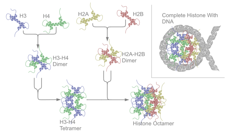
A deoxyribonucleoprotein (DNP) is a complex of DNA and protein.[2] The prototypical examples are nucleosomes, complexes in which genomic DNA is wrapped around clusters of eight histone proteins in eukaryotic cell nuclei to form chromatin. Protamines replace histones during spermatogenesis.
The most widespread deoxyribonucleoproteins are nucleosomes, in which the component is nuclear DNA. The proteins combined with DNA are histones and protamines; the resulting nucleoproteins are located in chromosomes. Thus, the entire chromosome, i.e. chromatin in eukaryotes consists of such nucleoproteins.[3][4]
Viruses
Many viruses are little more than an organized collection of deoxyribonucleoproteins.
Recombination intermediates
Homologous recombination is a process for repairing DNA that appears to be nearly universal. A central intermediate step in this process is the interaction of multiple copies of a recombinase protein with single-stranded DNA to form a DNP filament. Recombinases employed in this process are produced by archaea (RadA recombinase)[5], by bacteria (RecA recombinase)[6] and by eukaryotes from yeast to humans (Rad51 and Dmc1 recombinases).[7]
Ribonucleoproteins
A ribonucleoprotein (RNP) is a complex of ribonucleic acid and RNA-binding protein. These complexes play an integral part in a number of important biological functions that include DNA replication, regulating gene expression[9] and regulating the metabolism of RNA.[10] A few examples of RNPs include the ribosome, the enzyme telomerase, vault ribonucleoproteins, RNase P, hnRNP and small nuclear RNPs (snRNPs), which have been implicated in pre-mRNA splicing (spliceosome) and are among the main components of the nucleolus.[11]Some viruses are simple ribonucleoproteins, containing only one molecule of RNA and a number of identical protein molecules. Others are ribonucleoprotein or deoxyribonucleoprotein complexes containing a number of different proteins, and exceptionally more nucleic acid molecules. Currently, over 2000 RNPs can be found in the RCSB Protein Data Bank (PDB).[12] Furthermore, the Protein-RNA Interface Data Base (PRIDB) possesses a collection of information on RNA-protein interfaces based on data drawn from the PDB.[13] Some common features of protein-RNA interfaces were deduced based on known structures. For example, RNP in snRNPs have an RNA-binding motif in its RNA-binding protein. Aromatic amino acid residues in this motif result in stacking interactions with RNA. Lysine residues in the helical portion of RNA-binding proteins help to stabilize interactions with nucleic acids. This nucleic acid binding is strengthened by electrostatic attraction between the positive lysine side chains and the negative nucleic acid phosphate backbones. Additionally, it is possible to model RNPs computationally.[14] Although computational methods of deducing RNP structures are less accurate than experimental methods, they provide a rough model of the structure which allows for predictions of the identity of significant amino acids and nucleotide residues. Such information helps in understanding the overall function the RNP.
'RNP' can also refer to ribonucleoprotein particles. Ribonucleoprotein particles are distinct intracellular foci for post-transcriptional regulation. These particles play an important role in influenza A virus replication.[16] The influenza viral genome is composed of eight ribonucleoprotein particles formed by a complex of negative-sense RNA bound to a viral nucleoprotein. Each RNP carries with it an RNA polymerase complex. When the nucleoprotein binds to the viral RNA, it is able to expose the nucleotide bases which allow the viral polymerase to transcribe RNA. At this point, once the virus enters a host cell it will be prepared to begin the process of replication.
Anti-RNP antibodies
Anti-RNP antibodies are autoantibodies associated with mixed connective tissue disease and are also detected in nearly 40% of Lupus erythematosus patients. Two types of anti-RNP antibodies are closely related to Sjögren's syndrome: SS-A (Ro) and SS-B (La). Autoantibodies against snRNP are called Anti-Smith antibodies and are specific for SLE. The presence of a significant level of anti-U1-RNP also serves a possible indicator of MCTD when detected in conjunction with several other factors.[17]
Functions
In eukaryotic cells, DNA is associated with about an equal mass of histone proteins in a highly condensed nucleoprotein complex called chromatin.[18] Deoxyribonucleoproteins in this kind of complex interact to generate a multiprotein regulatory complex in which the intervening DNA is looped or wound. The deoxyribonucleoproteins participate in regulating DNA replication and transcription.[19]
The ribonucleoproteins play a role of protection. mRNAs never occur as free RNA molecules in the cell. They always associate with ribonucleoproteins and function as ribonucleoprotein complexes.[18]
In the same way, the genomes of negative-strand RNA viruses never exist as free RNA molecule. The ribonucleoproteins protect their genomes from RNase.[20] Nucleoproteins are often the major antigens for viruses because they have strain-specific and group-specific antigenic determinants.
Structure

Through crystallographic methods, the specific spatial structure and biological functions of many nucleoproteins are understood.[21][22] The structures of many viral nucleoproteins have been determined, including those of influenza,[23] rabies,[24] Ebola, Bunyamwera,[25] Schmallenberg,[25] Hazara,[26] Crimean-Congo hemorrhagic fever[27] and Lassa.[28] Important techniques for detecting the structures of nucleoproteins include X-ray diffraction, nuclear magnetic resonance and cryo-electron microscopy.
See also
References
- ^ Nucleoproteins at the U.S. National Library of Medicine Medical Subject Headings (MeSH)
- ^ Deoxyribonucleoproteins at the U.S. National Library of Medicine Medical Subject Headings (MeSH)
- ^ Graeme K. Hunter G. K. (2000): Vital Forces. The discovery of the molecular basis of life. Academic Press, London 2000, ISBN 0-12-361811-8.
- ^ Nelson D. L., Michael M. Cox M. M. (2013): Lehninger Principles of Biochemistry. W. H. Freeman, ISBN 978-1-4641-0962-1.
- ^ Seitz EM, Brockman JP, Sandler SJ, Clark AJ, Kowalczykowski SC (1998). "RadA protein is an archaeal RecA protein homolog that catalyzes DNA strand exchange". Genes Dev. 12 (9): 1248–53. PMC 316774. PMID 9573041.
- ^ Cox MM, Goodman MF, Kreuzer KN, Sherratt DJ, Sandler SJ, Marians KJ (2000). "The importance of repairing stalled replication forks". Nature. 404 (6773): 37–41. doi:10.1038/35003501. PMID 10716434.
- ^ Crickard JB, Kaniecki K, Kwon Y, Sung P, Greene EC (2018). "Spontaneous self-segregation of Rad51 and Dmc1 DNA recombinases within mixed recombinase filaments". J. Biol. Chem. doi:10.1074/jbc.RA117.001143. PMID 29382724.
{{cite journal}}: CS1 maint: unflagged free DOI (link) - ^ Muller, Mandy; Hutin, Stephanie; Marigold, Oliver; Li, Kathy H.; Burlingame, Al; Glaunsinger, Britt A. (2015-05-12). "A Ribonucleoprotein Complex Protects the Interleukin-6 mRNA from Degradation by Distinct Herpesviral Endonucleases". PLoS Pathogens. 11 (5): e1004899. doi:10.1371/journal.ppat.1004899. ISSN 1553-7366. PMC 4428876. PMID 25965334.
{{cite journal}}: CS1 maint: unflagged free DOI (link) - ^ Hogan, Daniel J; Riordan, Daniel P; Gerber, André P; Herschlag, Daniel; Brown, Patrick O (2016-11-07). "Diverse RNA-Binding Proteins Interact with Functionally Related Sets of RNAs, Suggesting an Extensive Regulatory System". PLoS Biology. 6 (10): e255. doi:10.1371/journal.pbio.0060255. ISSN 1544-9173. PMC 2573929. PMID 18959479.
{{cite journal}}: CS1 maint: unflagged free DOI (link) - ^ Lukong, Kiven E.; Chang, Kai-wei; Khandjian, Edouard W.; Richard, Stéphane (2008-08-01). "RNA-binding proteins in human genetic disease". Trends in genetics: TIG. 24 (8): 416–425. doi:10.1016/j.tig.2008.05.004. ISSN 0168-9525. PMID 18597886.
- ^ "Ribonucleoprotein". www.uniprot.org. Retrieved 2016-11-07.
- ^ Bank, RCSB Protein Data. "RCSB Protein Data Bank - RCSB PDB".
{{cite journal}}: Cite journal requires|journal=(help) - ^ Lewis, Benjamin A.; Walia, Rasna R.; Terribilini, Michael; Ferguson, Jeff; Zheng, Charles; Honavar, Vasant; Dobbs, Drena (2016-11-07). "PRIDB: a protein–RNA interface database". Nucleic Acids Research. 39 (Database issue): D277–D282. doi:10.1093/nar/gkq1108. ISSN 0305-1048. PMC 3013700. PMID 21071426.
- ^ Tuszynska, Irina; Matelska, Dorota; Magnus, Marcin; Chojnowski, Grzegorz; Kasprzak, Joanna M.; Kozlowski, Lukasz P.; Dunin-Horkawicz, Stanislaw; Bujnicki, Janusz M. (2014-02-01). "Computational modeling of protein-RNA complex structures". Methods (San Diego, Calif.). 65 (3): 310–319. doi:10.1016/j.ymeth.2013.09.014. ISSN 1095-9130. PMID 24083976.
- ^ Momose, Fumitaka; Sekimoto, Tetsuya; Ohkura, Takashi; Jo, Shuichi; Kawaguchi, Atsushi; Nagata, Kyosuke; Morikawa, Yuko (2011-06-22). "Apical Transport of Influenza A Virus Ribonucleoprotein Requires Rab11-positive Recycling Endosome". PLoS ONE. 6 (6): e21123. doi:10.1371/journal.pone.0021123. ISSN 1932-6203. PMC 3120830. PMID 21731653.
{{cite journal}}: CS1 maint: unflagged free DOI (link) - ^ Baudin, F; Bach, C; Cusack, S; Ruigrok, R W (1994-07-01). "Structure of influenza virus RNP. I. Influenza virus nucleoprotein melts secondary structure in panhandle RNA and exposes the bases to the solvent". The EMBO Journal. 13 (13): 3158–3165. ISSN 0261-4189. PMC 395207. PMID 8039508.
- ^ "Mixed Connective Tissue Disease (MCTD) | Cleveland Clinic". my.clevelandclinic.org. Retrieved 2016-11-07.
- ^ a b Lodish, Harvey. Molecular Cell Biology.
- ^ Echols, Harrison (1990). "Nucleoprotein structures initiating DNA replication, transcription, and site-specific recombination". The Journal of Biological Chemistry. 265: 14697–700. PMID 2203758.
- ^ Ruigrok, Rob WH; Crépin, Thibaut; Kolakofsky, Dan. "Nucleoproteins and nucleocapsids of negative-strand RNA viruses". Current Opinion in Microbiology. 14 (4): 504–510. doi:10.1016/j.mib.2011.07.011.
- ^ Graeme K. Hunter G. K. (2000): Vital Forces. The discovery of the molecular basis of life. Academic Press, London 2000, ISBN 0-12-361811-8.
- ^ Nelson D. L., Cox M. M. (2013): Lehninger Biochemie. Springer, ISBN 978-3-540-68637-8.
- ^ Ng, Andy Ka-Leung; Wang, Jia-Huai; Shaw, Pang-Chui (2009-05-27). "Structure and sequence analysis of influenza A virus nucleoprotein". Science in China Series C: Life Sciences. 52 (5): 439–449. doi:10.1007/s11427-009-0064-x. ISSN 1006-9305.
- ^ Albertini, Aurélie A. V.; Wernimont, Amy K.; Muziol, Tadeusz; Ravelli, Raimond B. G.; Clapier, Cedric R.; Schoehn, Guy; Weissenhorn, Winfried; Ruigrok, Rob W. H. (2006-07-21). "Crystal Structure of the Rabies Virus Nucleoprotein-RNA Complex". Science. 313 (5785): 360–363. doi:10.1126/science.1125280. ISSN 0036-8075. PMID 16778023.
- ^ a b Ariza, A.; Tanner, S. J.; Walter, C. T.; Dent, K. C.; Shepherd, D. A.; Wu, W.; Matthews, S. V.; Hiscox, J. A.; Green, T. J. (2013-06-01). "Nucleocapsid protein structures from orthobunyaviruses reveal insight into ribonucleoprotein architecture and RNA polymerization". Nucleic Acids Research. 41 (11): 5912–5926. doi:10.1093/nar/gkt268. ISSN 0305-1048. PMC 3675483. PMID 23595147.
- ^ Surtees, Rebecca; Ariza, Antonio; Punch, Emma K.; Trinh, Chi H.; Dowall, Stuart D.; Hewson, Roger; Hiscox, Julian A.; Barr, John N.; Edwards, Thomas A. (2015-01-01). "The crystal structure of the Hazara virus nucleocapsid protein". BMC Structural Biology. 15: 24. doi:10.1186/s12900-015-0051-3. ISSN 1472-6807. PMC 4696240. PMID 26715309.
{{cite journal}}: CS1 maint: unflagged free DOI (link) - ^ Carter, Stephen D.; Surtees, Rebecca; Walter, Cheryl T.; Ariza, Antonio; Bergeron, Éric; Nichol, Stuart T.; Hiscox, Julian A.; Edwards, Thomas A.; Barr, John N. (2012-10-15). "Structure, Function, and Evolution of the Crimean-Congo Hemorrhagic Fever Virus Nucleocapsid Protein". Journal of Virology. 86 (20): 10914–10923. doi:10.1128/JVI.01555-12. ISSN 0022-538X. PMC 3457148. PMID 22875964.
- ^ Qi, Xiaoxuan; Lan, Shuiyun; Wang, Wenjian; Schelde, Lisa McLay; Dong, Haohao; Wallat, Gregor D.; Ly, Hinh; Liang, Yuying; Dong, Changjiang. "Cap binding and immune evasion revealed by Lassa nucleoprotein structure". Nature. 468 (7325): 779–783. doi:10.1038/nature09605. PMC 3057469. PMID 21085117.
External links
This cell biology article is a stub. You can help Wikipedia by expanding it. |